Why a Kidney Stone Starts as Invisible Crystals
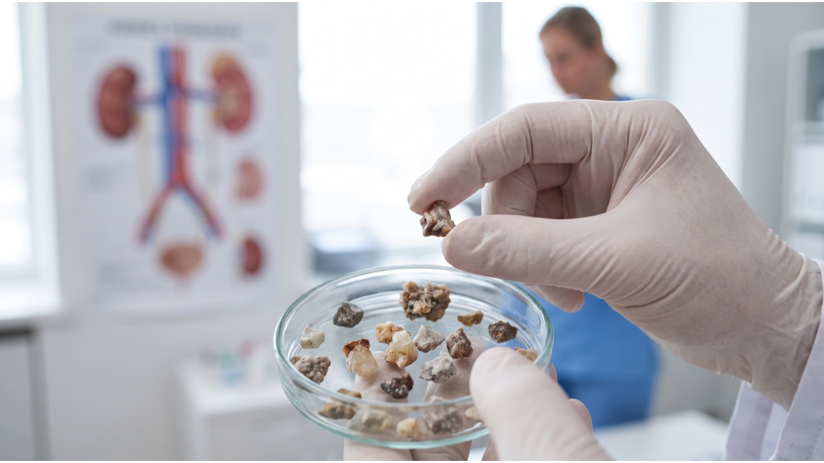
Few common medical events are described in language as dramatic as a kidney stone, and the comparison to childbirth pain is not exaggeration for many patients. Yet the process that produces this agony begins almost invisibly, as microscopic mineral crystals form silently inside the kidney long before anyone feels a thing, growing undetected for weeks or months before a stone is large enough to cause a problem.
The kidneys constantly filter waste products and excess minerals out of the blood into urine, a process that under normal circumstances keeps these substances dissolved and harmlessly flushed out of the body. A kidney stone forms when certain minerals in urine become concentrated enough that they can no longer stay dissolved and instead begin crystallizing out of solution.
These initial crystals are far too small to be felt or even seen without magnification, and in most people they are simply flushed out with normal urine flow before they can grow any larger, which is why the vast majority of people who form microscopic crystals never go on to develop a symptomatic stone at all.
How Supersaturation Actually Triggers Crystal Growth
The chemical concept underlying stone formation is called supersaturation, a state in which urine contains more of a dissolved mineral than the surrounding fluid can actually hold in solution, similar to the way sugar stirred into tea eventually stops dissolving and begins settling as visible grains once the liquid reaches its saturation point.
Urine is a genuinely complex chemical solution containing calcium, oxalate, uric acid, phosphate, and various other compounds, all held in a delicate balance influenced by fluid intake, diet, urine pH, and the presence of natural inhibitor substances like citrate that actively work to keep these minerals dissolved.
When that balance tips toward supersaturation, whether from excessive mineral excretion, insufficient fluid to dilute the urine, or a drop in protective inhibitors, tiny crystals begin to nucleate and then progressively attract more dissolved mineral, growing layer by layer over time into a solid mass.
Why Calcium Oxalate Is the Most Common Stone Type
The large majority of kidney stones are composed primarily of calcium oxalate, formed when calcium and oxalate, both normally present in urine at manageable levels, bind together and crystallize once their combined concentration exceeds what the urine can hold in solution.
Oxalate is a naturally occurring compound found in many otherwise healthy foods, including spinach, nuts, and chocolate, and while dietary oxalate contributes to urinary oxalate levels, the body's own metabolism also produces a meaningful share, meaning oxalate intake alone does not fully explain who forms stones and who does not.
Genetics, chronic dehydration, certain digestive conditions that increase oxalate absorption, and diets high in animal protein and sodium all influence how much calcium and oxalate end up in urine at any given time, which is why calcium oxalate stone risk varies so substantially between individuals eating broadly similar diets.
How Uric Acid Stones Form Differently
Uric acid stones form through a distinct chemical pathway tied closely to urine acidity rather than mineral concentration alone. Uric acid becomes far less soluble in acidic urine, so people whose urine runs persistently acidic, often linked to diets very high in animal protein, are at meaningfully elevated risk regardless of how much uric acid they actually excrete.
This type is also strongly associated with conditions like gout, obesity, and type 2 diabetes, all of which can independently push urine toward the acidic range that favours uric acid crystallization, making uric acid stones something of a metabolic marker as much as a standalone urinary problem.
Because the underlying mechanism is chemical rather than purely a matter of mineral excess, uric acid stones can sometimes be dissolved with medication that alkalinizes urine, an option not available for calcium-based stones once they have fully formed.
Why Struvite and Cystine Stones Are Rarer But Distinct
Struvite stones form through an entirely different mechanism involving certain bacteria that produce an enzyme capable of splitting urea into ammonia, which sharply raises urine pH and creates ideal chemical conditions for these particular minerals to crystallize rapidly, sometimes forming large branching stones within weeks of a urinary tract infection.
Cystine stones are rarer still, caused by a specific inherited genetic condition that prevents the kidneys from properly reabsorbing the amino acid cystine, allowing it to accumulate in urine at levels that inevitably exceed its very limited solubility regardless of diet or hydration habits.
Both of these less common types generally require different management approaches than the calcium and uric acid stones that make up the overwhelming majority of cases, underscoring why accurate stone composition analysis matters for choosing effective long-term prevention.
What Actually Causes the Severe Pain
The pain associated with kidney stones, often described as among the most intense a person can experience, comes not from the stone sitting quietly inside the kidney but from what happens once it begins moving into the narrow ureter, the tube connecting kidney to bladder.
A stone lodged in the ureter partially or completely blocks the normal flow of urine, causing urine to back up and the kidney itself to swell, a condition called hydronephrosis, while the ureter simultaneously spasms in painful waves as smooth muscle tries unsuccessfully to push the obstruction through.
This combination of organ swelling and forceful muscular spasm produces the classic pattern of severe, wave-like flank pain that can radiate toward the groin as the stone migrates lower, frequently accompanied by nausea, restlessness, and an inability to find a comfortable position, all reflecting genuine visceral distress rather than exaggeration.
How Stone Size Determines Whether It Passes on Its Own
Stone size is the single strongest predictor of whether natural passage is realistic. Stones smaller than roughly four millimetres pass spontaneously in the large majority of cases, often within a couple of weeks with adequate hydration and pain control, while stones in the intermediate range require closer monitoring.
Stones larger than about ten millimetres are unlikely to pass without intervention and carry a higher risk of causing prolonged obstruction, significant kidney swelling, or infection if left untreated, which is why doctors typically recommend active removal for stones above this threshold rather than waiting.
Location within the urinary tract also matters considerably, since a stone lodged near the narrower points of the ureter, particularly close to where it enters the bladder, is more likely to become genuinely stuck than one still travelling through wider sections of the passage.
Why Dehydration Is the Single Biggest Risk Factor
Insufficient fluid intake is consistently identified as the most significant modifiable risk factor across essentially all stone types, because concentrated urine directly drives the supersaturation process that initiates crystal formation in the first place.
People living in hot climates, athletes who lose substantial fluid through sweat, and anyone with habitually low water intake are disproportionately represented among stone formers, a pattern documented consistently enough that urologists in warm-climate regions actively emphasize hydration counselling as frontline prevention.
The protective effect is genuinely dose-dependent: studies tracking urine output have found that producing a larger daily volume of dilute urine correlates directly with lower stone recurrence, making sustained daily fluid intake one of the few prevention measures with unambiguous, broadly applicable evidence behind it.
How Diet Genuinely Affects Stone Risk
Popular advice to simply cut calcium intake for calcium stone prevention is, somewhat counterintuitively, often wrong. Dietary calcium normally binds oxalate together in the digestive tract before either is absorbed, and too little calcium can actually leave more free oxalate available for absorption and eventual excretion in urine.
Reducing sodium intake tends to matter considerably more, since high sodium increases urinary calcium excretion independent of dietary calcium itself, and reducing animal protein similarly lowers both calcium and uric acid excretion while also raising protective urinary citrate levels.
For people who have already formed a stone, a formal metabolic evaluation analysing 24-hour urine composition is generally more useful than generic dietary advice, since the specific mineral imbalance driving one person's stones can differ meaningfully from another's despite similar diets.
How Doctors Actually Diagnose a Kidney Stone
A non-contrast CT scan of the abdomen and pelvis is the most accurate diagnostic tool, capable of detecting stones of nearly any composition and size while also revealing the degree of obstruction and kidney swelling present, information that directly shapes treatment decisions.
Ultrasound is frequently used as a lower-radiation alternative, particularly for pregnant patients or for routine follow-up, though it is somewhat less sensitive for detecting smaller stones compared to CT imaging, and simple urine and blood tests help identify infection, kidney function changes, and clues about likely stone composition.
When a stone is eventually passed or surgically removed, sending it for laboratory composition analysis is considered essential rather than optional, since the specific mineral makeup directly determines which prevention strategy is actually likely to work for that individual patient.
What Happens When a Stone Will Not Pass Naturally
When pain becomes unmanageable, infection develops behind an obstructed stone, or kidney function is genuinely threatened by prolonged blockage, doctors will not wait for natural passage and instead move toward active intervention, sometimes urgently if infection is present alongside obstruction.
A temporary ureteral stent, a thin flexible tube placed to hold the ureter open and allow urine to drain around the stone, is frequently used as an immediate measure to relieve dangerous obstruction while a more definitive procedure is scheduled or while a stone is given additional time to pass on its own.
The choice between available definitive procedures depends heavily on stone size, location, and composition, with urologists weighing the relative invasiveness, effectiveness, and recovery time of each option against the specific clinical picture presented by that particular stone.
How Shockwave Lithotripsy Actually Breaks Up Stones
Extracorporeal shockwave lithotripsy directs focused shockwaves through the skin from outside the body, precisely targeted at the stone using imaging guidance, fracturing it into smaller fragments small enough to pass naturally through subsequent urination over the following days or weeks.
This approach is genuinely noninvasive, requiring no incisions, and works well for many moderate-sized stones in accessible locations, though it is less effective against very hard stone compositions like cystine and can require multiple sessions for larger or denser stones.
Ureteroscopy, an alternative approach involving a thin scope passed through the urinary tract itself to directly visualize and fragment or remove the stone using a laser, offers higher success rates for stones in certain locations and is often preferred when lithotripsy is unlikely to be fully effective.
Why Kidney Stones Recur So Often
Roughly half of people who form one kidney stone will form another within five to ten years without any change to diet or hydration habits, reflecting the fact that the underlying metabolic and dietary conditions that produced the first stone typically remain unchanged unless deliberately addressed.
This high recurrence rate is precisely why urologists increasingly emphasize prevention counselling as an essential part of stone treatment rather than an optional afterthought, since simply removing or passing one stone does nothing to correct whatever imbalance produced it in the first place.
Family history also plays a genuine role, with people who have a first-degree relative who formed stones facing meaningfully elevated personal risk, suggesting a real genetic contribution to how efficiently different individuals excrete and regulate the relevant urinary minerals.
Certain medical conditions and medications also raise recurrence risk independently of diet, including inflammatory bowel disease and prior gastric bypass surgery, both of which alter how the intestines absorb calcium and oxalate, as well as chronic dehydration linked to climate or occupation, which is one reason people working outdoors in consistently hot environments show elevated rates of stone formation regardless of what they eat.
What Genuinely Reduces Long-Term Recurrence Risk
Consistent, adequate fluid intake sufficient to produce a substantial daily urine volume remains the single most broadly effective prevention measure across nearly all stone types, and it requires no prescription, specialized testing, or significant lifestyle disruption to implement.
For patients with a confirmed pattern of recurrence, targeted dietary adjustments guided by actual 24-hour urine testing, and in some cases specific medications that alter urine chemistry or reduce mineral excretion, can meaningfully lower the odds of forming another stone compared to hydration alone.
What begins as an invisible chemical imbalance inside the kidney can escalate into one of medicine's most acutely painful common events, but understanding that pathway from crystal to stone to obstruction is exactly what makes modern prevention and treatment genuinely effective rather than a matter of simply waiting out the pain.
Sources
- Wikipedia — overview of kidney stone types, formation, and treatment
- National Institute of Diabetes and Digestive and Kidney Diseases (NIH) — clinical information on kidney stone causes and management
- World Health Organization — global health data relevant to urinary and metabolic disease
- American Urological Association — clinical guidelines for kidney stone diagnosis and treatment
- National Health Service (UK) — patient-facing guidance on kidney stone symptoms and care
FAQ
Why do kidney stones hurt so much?
The pain comes mainly from the ureter spasming and stretching as it tries to push a stone past an obstruction, combined with swelling of the kidney caused by trapped urine backing up behind the blockage.
Will a kidney stone always need surgery?
No — most small stones under about 4-5 millimetres pass on their own with hydration and pain management, while larger or stuck stones often require shockwave therapy, ureteroscopy, or other procedures.
Does drinking more water actually prevent kidney stones?
Yes — adequate hydration is one of the most consistently effective prevention strategies because it dilutes urine and reduces the concentration of stone-forming minerals below the threshold needed for crystals to form.
Is cutting out calcium the right way to prevent calcium stones?
No — counterintuitively, too little dietary calcium can actually increase stone risk for some people, because calcium normally binds oxalate in the gut; reducing sodium and animal protein is generally a more effective strategy.
Do kidney stones come back after treatment?
Recurrence is common — roughly half of people who form one stone will form another within several years without dietary or medical changes, which is why prevention strategies matter as much as treating the initial stone.
About the Author
We reference Wikipedia, the National Institute of Diabetes and Digestive and Kidney Diseases, the World Health Organization, the American Urological Association, and the UK National Health Service to explain the background and current understanding of this topic.
Loved This Article?
Share it on WhatsApp → Share it on WhatsApp
Get more guides in your inbox — Subscribe to our newsletter for weekly surprising stories from Egypt, Saudi Arabia, Dubai, and beyond.